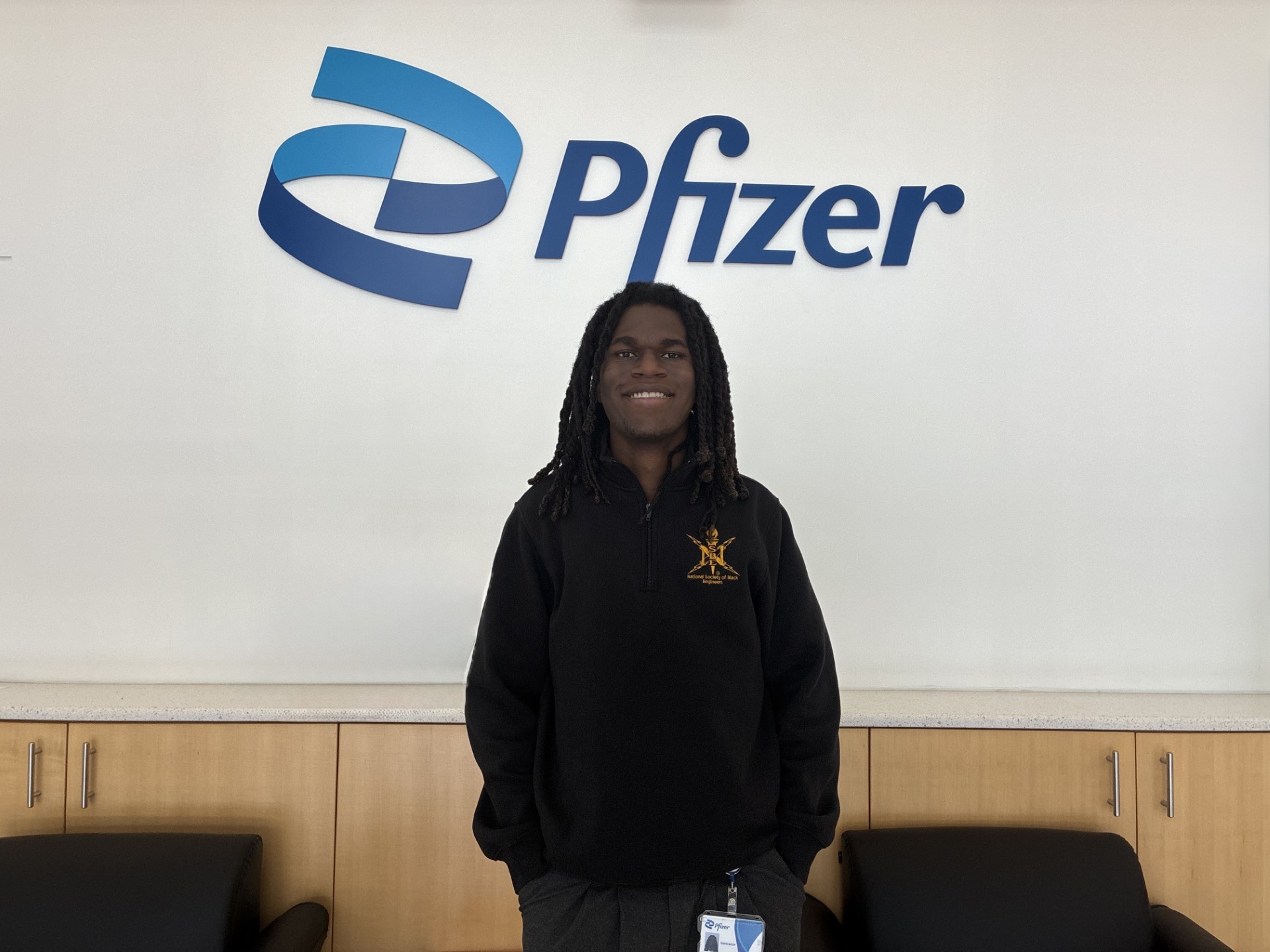
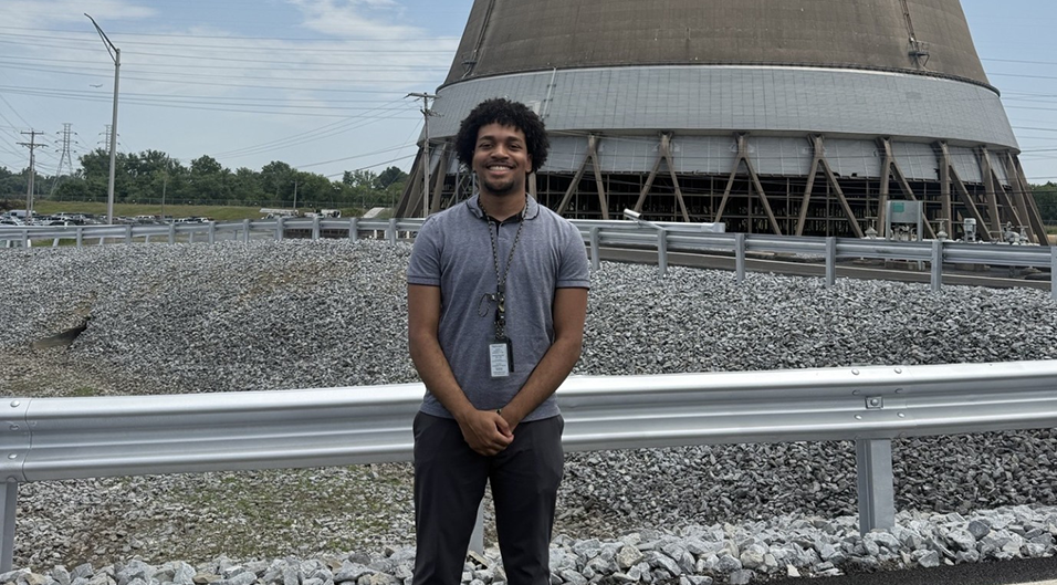
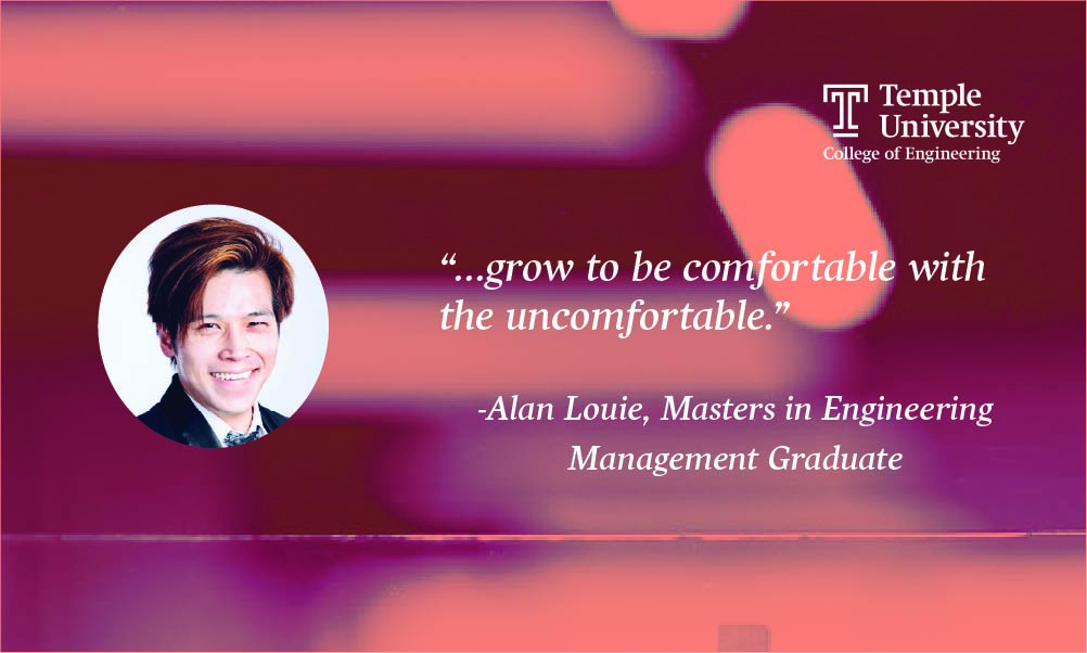
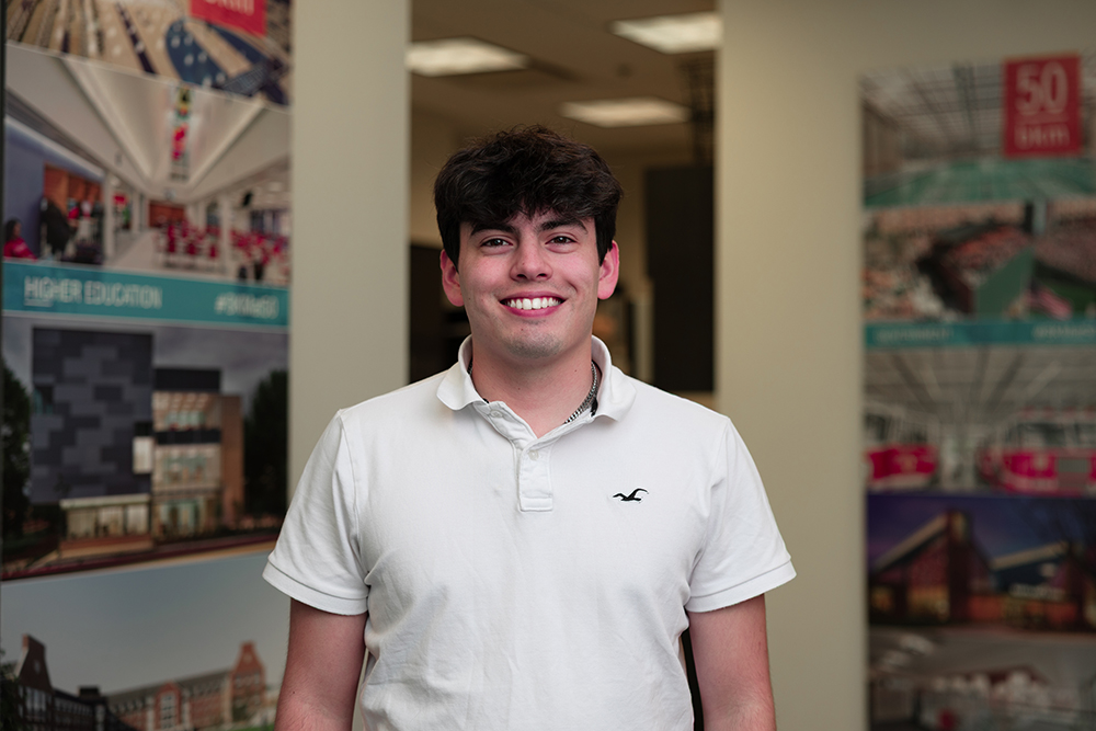
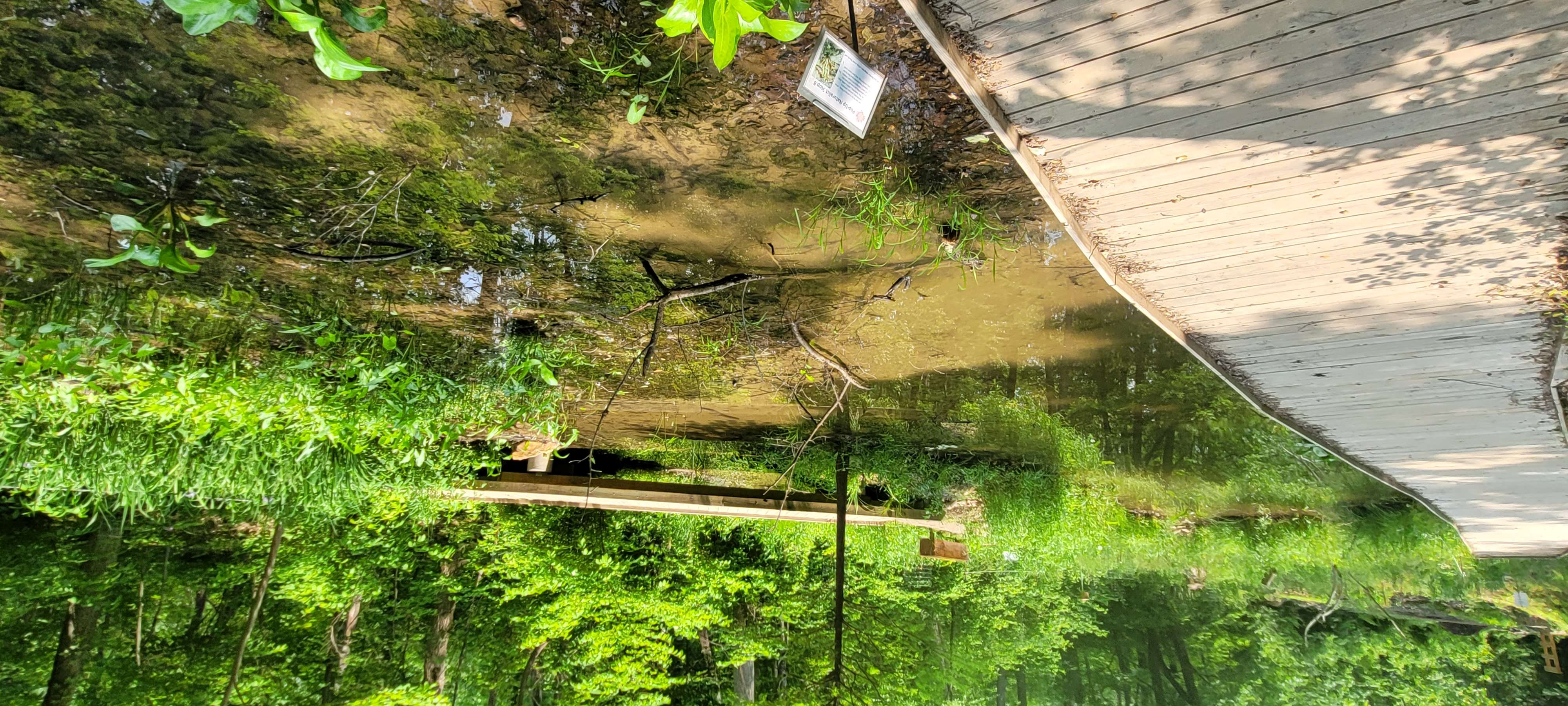
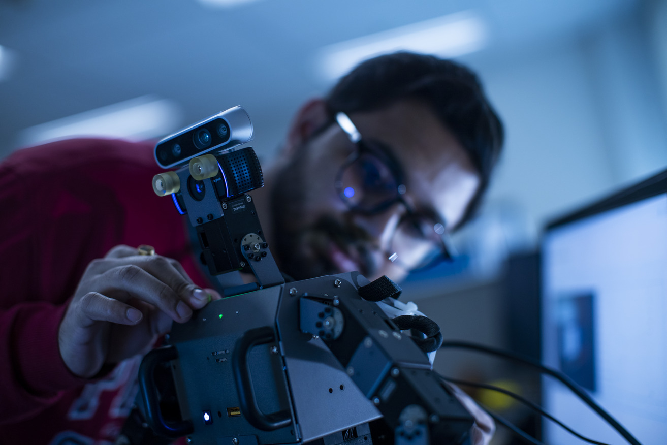
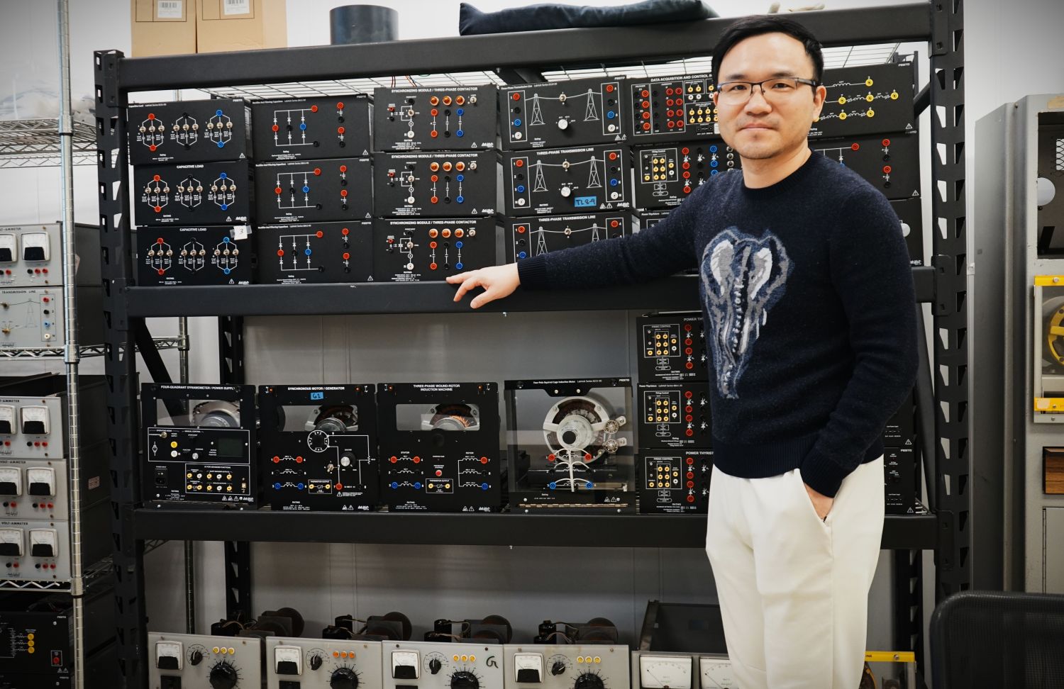
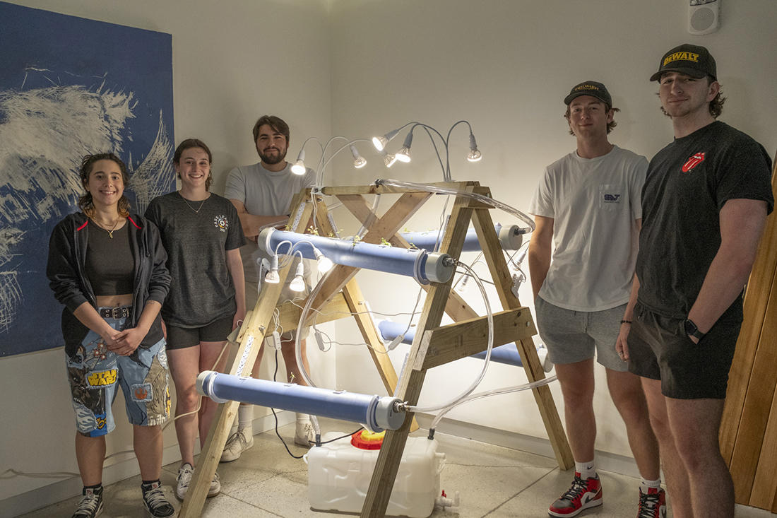
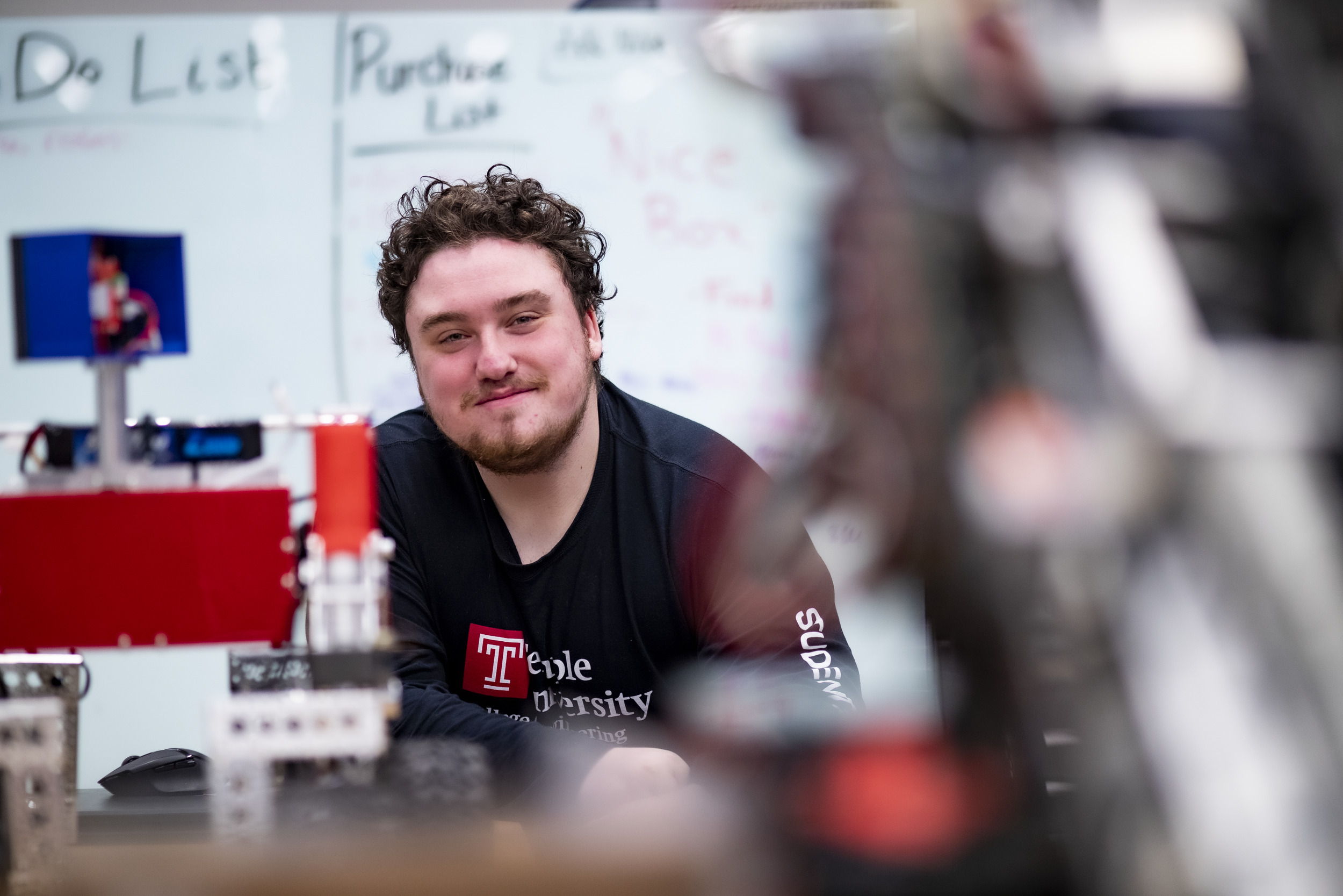
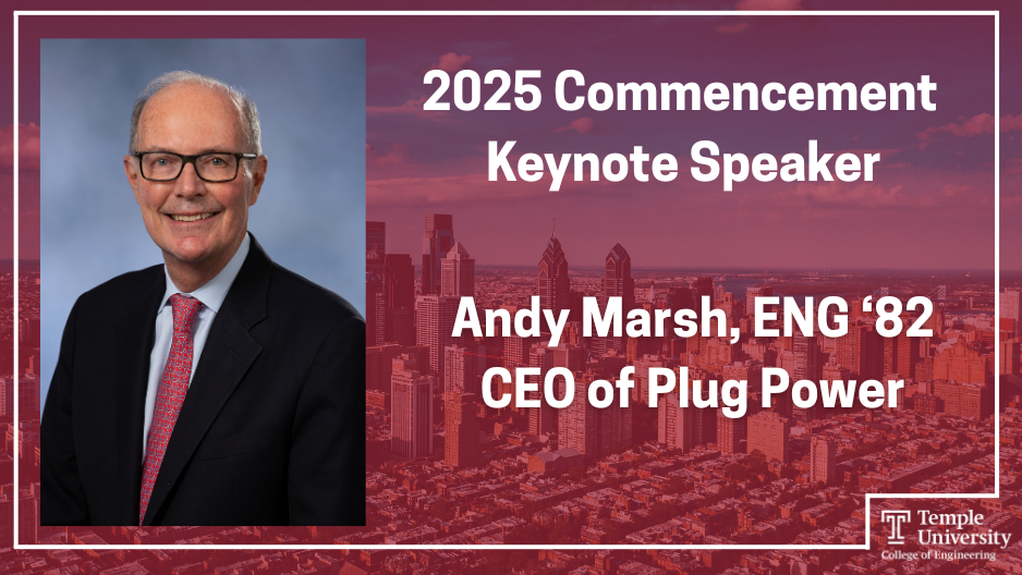
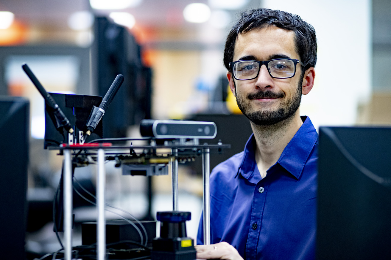
Photo by Adam Lawrence Jul. 30, 2025 Owls on the job: Adam Lawrence, Central Design Organization Engineering Intern By Casey TinneyThis summer, electrical engineering student Adam Lawrence is back at Constellation for his second […] Photo by Alan Louie Jul. 18, 2025 Alumni Profile: Alan Louie –How the Masters in Engineering Management changed his trajectory By Jesse ONeillAlan Louie is a Senior Project Team Lead and Technical Project Engineer at Blue Origin, a private […] Photo by Melissa Newman Jul. 9, 2025 Owls on the job: Javier Schorle, Electrical Intern By Casey TinneySummer break is often a time for students to relax, catch up on course work, and in many cases, […] Photo by Ryan S. Brandenberg Jul. 9, 2025 Bioengineering professor awarded inaugural Blue Sky Initiative grant By Casey TinneyAssistant Professor of Bioengineering Karin Wang, PhD, was recently awarded one of the inaugural […] Left: The Daisy Portable Bidet Logo, Right: Cheri MitchellPhoto by Provided by Cybil Seneker Jun. 23, 2025 Former senior design project seeks to increase autonomy for disabled individuals By Casey TinneyThe College of Engineering’s Senior Design Project is the culmination of an engineering student’s […] Wetlands 2025 in Full BloomPhoto by Kevin Magerr Jun. 12, 2025 One Year Later: An Ecosystem Thrives after Temple Engineers help Restore the Wetlands By Jesse ONeillA year after Temple University engineering students and environmental partners completed the […] Photo by Provided by Aysia Davis Jun. 6, 2025 Career fair connection turns into full time job offer By Casey TinneyIn addition to developing strong technical skills in the classroom, the College of Engineering […] Photo by Ryan S. Brandenberg May 28, 2025 The Cycle of Kindness: A Story of Philanthropy By The College of EngineeringPhilanthropy doesn't start with money. It begins with a simple act of kindness. Professor Emeritus […] Photo by Casey Tinney May 19, 2025 Engineering professor explains potential causes and outcomes of power blackouts By Casey TinneyOn Monday, April 28, 2025, residents on the Iberian Peninsula, specifically in Spain and Portugal, […] Photo by Casey Tinney May 19, 2025 Engineering professor explains potential causes and outcomes of power blackouts By Casey TinneyOn Monday, April 28, 2025, residents on the Iberian Peninsula, specifically in Spain and Portugal, […] Photo by Provided by Greg and Michele Kelly May 12, 2025 Building Bridges of Opportunity: The Greg and Michele Kelly Story By Casey TinneyA chance meeting at a Temple football game blossomed into 47 years of partnership, success, and […] Engineers for Climate Action members with the hydroponic system in Charles LibraryPhoto by Joseph V. Labolito Apr. 28, 2025 Engineers for Climate Action install hydroponic system for urban gardening on campus By Casey TinneyThe student organization Engineers for Climate Action’s sustainable hydroponic system will be on […] Photo by Ryan Brandenberg Apr. 17, 2025 2025 Student Graduation Speaker By Jesse ONeill and Jared LevinThe College of Engineering is honored to introduce this year’s student graduation speaker, Jared […] Photo by Photo provided by Andy Marsh Apr. 14, 2025 2025 Graduation Keynote Speaker: Andy Marsh By Jesse ONeillOn May 8th 2025, Andy Marsh, a proud Temple Owl, will return to the nest to impart his wisdom to […] Photo by Ryan S. Brandenberg Apr. 11, 2025 Philip Dames, PhD announced as Lindback Distinguished Teaching Award recipient By Casey TinneyPhilip Dames, PhD was named as a 2025 recipient of the Lindback Distinguished Teaching Award. Dames […] Load More
Photo by Adam Lawrence Jul. 30, 2025 Owls on the job: Adam Lawrence, Central Design Organization Engineering Intern By Casey TinneyThis summer, electrical engineering student Adam Lawrence is back at Constellation for his second […] Photo by Alan Louie Jul. 18, 2025 Alumni Profile: Alan Louie –How the Masters in Engineering Management changed his trajectory By Jesse ONeillAlan Louie is a Senior Project Team Lead and Technical Project Engineer at Blue Origin, a private […] Photo by Melissa Newman Jul. 9, 2025 Owls on the job: Javier Schorle, Electrical Intern By Casey TinneySummer break is often a time for students to relax, catch up on course work, and in many cases, […] Photo by Ryan S. Brandenberg Jul. 9, 2025 Bioengineering professor awarded inaugural Blue Sky Initiative grant By Casey TinneyAssistant Professor of Bioengineering Karin Wang, PhD, was recently awarded one of the inaugural […] Left: The Daisy Portable Bidet Logo, Right: Cheri MitchellPhoto by Provided by Cybil Seneker Jun. 23, 2025 Former senior design project seeks to increase autonomy for disabled individuals By Casey TinneyThe College of Engineering’s Senior Design Project is the culmination of an engineering student’s […] Wetlands 2025 in Full BloomPhoto by Kevin Magerr Jun. 12, 2025 One Year Later: An Ecosystem Thrives after Temple Engineers help Restore the Wetlands By Jesse ONeillA year after Temple University engineering students and environmental partners completed the […] Photo by Provided by Aysia Davis Jun. 6, 2025 Career fair connection turns into full time job offer By Casey TinneyIn addition to developing strong technical skills in the classroom, the College of Engineering […] Photo by Ryan S. Brandenberg May 28, 2025 The Cycle of Kindness: A Story of Philanthropy By The College of EngineeringPhilanthropy doesn't start with money. It begins with a simple act of kindness. Professor Emeritus […] Photo by Casey Tinney May 19, 2025 Engineering professor explains potential causes and outcomes of power blackouts By Casey TinneyOn Monday, April 28, 2025, residents on the Iberian Peninsula, specifically in Spain and Portugal, […] Photo by Casey Tinney May 19, 2025 Engineering professor explains potential causes and outcomes of power blackouts By Casey TinneyOn Monday, April 28, 2025, residents on the Iberian Peninsula, specifically in Spain and Portugal, […] Photo by Provided by Greg and Michele Kelly May 12, 2025 Building Bridges of Opportunity: The Greg and Michele Kelly Story By Casey TinneyA chance meeting at a Temple football game blossomed into 47 years of partnership, success, and […] Engineers for Climate Action members with the hydroponic system in Charles LibraryPhoto by Joseph V. Labolito Apr. 28, 2025 Engineers for Climate Action install hydroponic system for urban gardening on campus By Casey TinneyThe student organization Engineers for Climate Action’s sustainable hydroponic system will be on […] Photo by Ryan Brandenberg Apr. 17, 2025 2025 Student Graduation Speaker By Jesse ONeill and Jared LevinThe College of Engineering is honored to introduce this year’s student graduation speaker, Jared […] Photo by Photo provided by Andy Marsh Apr. 14, 2025 2025 Graduation Keynote Speaker: Andy Marsh By Jesse ONeillOn May 8th 2025, Andy Marsh, a proud Temple Owl, will return to the nest to impart his wisdom to […] Photo by Ryan S. Brandenberg Apr. 11, 2025 Philip Dames, PhD announced as Lindback Distinguished Teaching Award recipient By Casey TinneyPhilip Dames, PhD was named as a 2025 recipient of the Lindback Distinguished Teaching Award. Dames […]