Dr. Nancy Pleshko is using her experience with spectroscopy to help solve a problem affecting 30 million Americans.
For Dr. Nancy Pleshko, Professor of Bioengineering, spectroscopy has been a focal point going back to her doctoral work. Now, through funding secured from the National Institutes of Health (NIH), Dr. Pleshko is applying spectroscopic imaging to help Americans suffering from osteoarthritis in their knees and hips.
In this condition, the cartilage around the joint wears away and the tissue does not regenerate. Patients deal with increasing pain while performing everyday activities. The most common solution is knee or hip replacement using an artificial metal joint.
Doctors and researchers are trying to find a way to replace the worn away cartilage, instead of replacing the worn knee joint. There are ways to recreate the materials in a lab through tissue engineering. The catch seems to be when to harvest the materials and introduce them to the affected knee. Dr. Pleshko's research is applying her expertise in spectroscopic imaging techniques to judge when the engineered cultures are ready to implant.
Dr. Pleshko's lab is focused on ways to evaluate the development of the engineered tissue without destroying it. Current standard assessment procedures take samples of a growth batch to determine if the tissue is ready, which makes that tissue no longer able to develop further or be used for implants.
"The assumption is that you assess tissue A, and you're going to assume that tissue B that was growing in the same conditions is at the exact same level of maturity," Dr. Pleshko said. "Then you can implant tissue B based on the characteristics of tissue A, as that was grown in the same batch. We're just finding out more and more that's really not necessarily the case. There's so much variability that just arises from the process that's used to actually make the engineered tissues. No two constructs are alike and no two constructs mature at the same rate and have the exact same composition. There's a lot of variability."
The pre-clinical phase of the grant funding will begin in 2018, running for two years. She hopes the spectroscopic data collection during that period will help confirm the methodology and can be translated to eventual clinical application to implantation of engineered tissues.
Dr. Pleshko got her start in bioengineering with her doctoral work on spectroscopy at Rutgers University. She also spent years at the Hospital for Special Surgery in New York City as a researcher before coming to Temple in 2009 to work in the Mechanical Engineering department. In 2012, she was a founding faculty member in the new Bioengineering Department.
Throughout her career, she has been funded by the NIH, primarily through the National Institute on Arthritis and Musculoskeletal and Skin Diseases. On this project, she is working with Dr. Robert L. Mauck, working out of the University of Pennsylvania Perelman School of Medicine in the McKay Orthopaedic Research Lab.
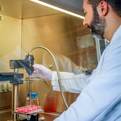
Dr. Pleshko can draw on clinicians and interns from Temple's College of Medicine, and finds her lab full of engineering students, including many undergraduates with medical aspirations. To better collect data on tissue development, a Senior Design team built a pressure sensor for a fiber optic probe that kept load constant on the examining probe used to take spectral images of the tissue. Other portions of the research project utilize different majors at the College of Engineering and the College of Science and Technology.
"It's really great to have the student involvement," she said. "There's many pre-med students at Temple, whether in biology or bioengineering. They look at a project like this and it's really exciting to them. It's some engineering, it's biology. It has clinical impact."